Hybridization assays: an interview with Helen Legakis

In this interview, Helen Legakis goes into detail about hybridization assays as she discusses how they can help to support PK and TK studies and what considerations to take into account when developing hybridization assays.
 Helen Legakis
Principal Scientist
Charles River Laboratories (Montreal, Canada)
Helen Legakis, Principal Scientist, oversees the oligonucleotide hybridization-ELISA services within the Immunochemistry department at Charles River’s site in Montreal, Canada.
She received her BSc in Biochemistry at McGill University (Montreal, Canada) in 2003 and throughout her graduate studies, she applied various immunobiological methods to determine the intracellular signaling pathway of a G-protein-coupled receptor linked to obesity.
In 2005, Helen completed her MSc in Biochemistry at McGill University and joined Charles River that same year where she was involved in developing, validating and coordinating bioanalytical drug programs for several pharmaceutical and biotechnology sectors in support of regulatory preclinical and clinical studies.
Helen works on various drug programs that include siRNA and antisense oligonucleotides, focusing on establishing hybridization methods for the quantitation of these oligonucleotides in various biological fluids and tissues.
- What are hybridization assays?
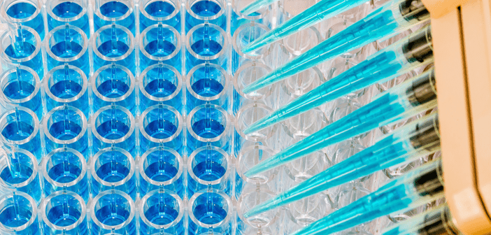
Hybridization-based assays are enzyme-linked immunosorbent assays (ELISAs) that are routinely used to measure oligonucleotide drug therapies that are at least 18 nucleotides in length. This type of immunoassay can be conducted in a 96-well plate and uses Watson-Crick base pairing to a complementary probe sequence to immobilize and quantify the amount of oligonucleotide therapeutic present in a biological sample. Since complementary probes targeting the exact nucleobase sequence of the drug are used, minimal to no sample clean-up is needed and small volumes can be used to generate concentration results. These assays also provide enhanced assay sensitivity and sample throughput compared to conventional chromatographic techniques. Alternatively, the main limitation is assay specificity for an intact drug. Different hybridization-based formats have been developed to enhance assay specificity, particularly towards the 3’-end of the drug sequence where exonuclease activity predominantly occurs.
- How do hybridization assays help support PK and TK studies?
High-performance liquid chromatography (HPLC) and liquid chromatography-mass spectrometry (LC-MS) have been used for measuring large molecules in biological matrices. Although highly selective in distinguishing full-length drugs from generated catabolites, their insufficient sensitivity has limited their ability to fully characterize the pharmacokinetic profile of therapeutic oligonucleotides following systemic exposure. Assay sensitivity is particularly important during the terminal elimination phase when near complete biodistribution has occurred and very low levels of circulating drug are present in the blood. The increasing demand for ultra-sensitive bioanalytical assays has made hybridization-based ELISAs a highly desirable bioanalytical tool. A combination of bioanalytical platforms may be used to analyze drug concentrations in various biological matrices to fully characterize exposure-response and exposure-toxicity relationships.
- What steps can one take to optimize assay sensitivity with hybridization-ELISA techniques?
Assay sensitivity can be optimized to a drug program’s needs by enhancing the stringency of the hybridization reaction. This can be achieved by lowering the salt concentration of the reaction mixture and increasing the incubation temperature. The introduction of RNA analogues to the probe sequence can enhance the duplex Tm. Locked nucleic acids (LNAs), for example, contain a modified sugar moiety that enhances the probe Tm to a target sequence by approximately 4°C per insert. This enables the hybridization reaction to occur at more elevated temperatures, reducing duplexes formed from mismatched base pairings. Critical design must be used when incorporating LNAs into the probe sequence to avoid self-annealing that can result from this enhanced stability. Various detection systems such as colorimetric, fluorescent, or electrochemiluminescent output for example can also be evaluated to further enhance the signal-to-noise ratio of the method to establish the optimal lower limit of quantitation.
- What are some key considerations when developing and validating hybridization assays?
Similar to small molecules, oligonucleotide therapeutics are chemically synthesized and purified. Based on their size, they are considered large molecules and hybridization assays used for their analysis can be validated following FDA and ICH M10 guidance for large molecule analysis by ligand binding assays to support both pre-clinical and clinical filings. A certificate of analysis detailing purity and stability should be provided for each reference standard. The typical validation parameters tested for a bioanalytical method are selectivity, specificity, precision and accuracy, calibration curve range, prozone and dilution linearity. Another parameter is the assessment of stability to test sample handling, i.e., short-term stability following multiple rounds of freezing and thawing, bench-top stability at ambient room temperature and in a refrigerator set to maintain 4°C and prolonged storage stability. Once the bioanalytical method has been validated, the precision and accuracy of the assay are monitored during the in-study phase with the use of acceptance QC samples. Incurred sample reanalysis (ISR) also demonstrates whether the assay can accurately quantify drug concentrations in biological study samples.
- Can the same set of hybridization-ELISA probes be used to assess multiple therapeutic oligonucleotides?
Early-stage discovery programs can investigate multiple drug candidate sequences before selecting a final sequence. Depending on the assay format selected, multiple candidate sequences can be analyzed independently using a single set of assay probes if they share a common stretch of sequence homology. For example, a sandwich format uses complementary probes (capture and detection) that span approximately each half of the oligonucleotide sequence. In this scenario, the drug candidates must share sequence homology of at least 18 nucleotides to ensure that the assay probes can form a stable hybridized duplex and generate an assay response. Any modifications to the phosphodiester backbone and/or sugar moieties should not significantly impact hybridization efficiency if the nucleobase sequence in this homologous region is conserved across these candidate sequences. Once the working range is established in one candidate, the remaining sequences can be tested independently as a proof-of-concept to investigate whether the same working range can be applied.
This interview is a part of the Bioanalysis Zone Spotlight feature on the practical challenges of new modalities. For more expert opinions on this topic, visit our feature homepage
In association with:


