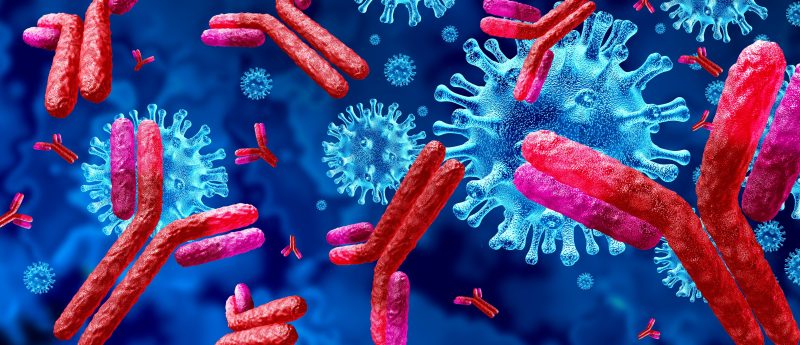
Utilizing monoclonal antibodies to regulate checkpoint proteins & boost immune response

Refer a colleague
Muromonab-CD3 was first approved in 1986 to help patients’ immune systems accept kidney transplants. Since then, the US FDA has approved more than 100 monoclonal antibodies (mAbs) to treat diseases ranging from autoimmune disorders, HIV, cancer and COVID-19. The first question scientists may ask when examining a new drug modality is whether it can interact with the target receptor and/or whether it will induce an immune response. To those ends, mAbs present several advantages and have become one of the most successful biotherapeutic drugs utilized in the treatments.
Compared to other drugs, monoclonal antibodies maintain a high affinity to their target antigen and demonstrate strong selectivity, specificity and sensitivity. Despite their extensive use, mAbs present several disadvantages, namely that they are very expensive and difficult to produce. Additionally, their large molecular size(~150 kDa) limits their tissue and tumor penetration, thus limiting their biodistribution and efficacy. ‘Humanized’ monoclonal antibodies mimic human antibody structures, which are close to naturally occurring Ab’s in humans. The humanized antibody may be unrecognized by (or at low risk of eliciting) anti-drug antibodies (ADAs) and neutralizing antibodies (NAbs). The ADAs and NAbs might otherwise inhibit the drug’s efficacy.
MAbs are also highly scalable and reproducible because they use hybrid cells (i.e., hybridoma) for antibody production. Hybridoma are gathered by injecting test articles with an antigen and then collecting an antibody-producing cell after an immune response occurs. This process provides a potentially unlimited source of identical monoclonal antibody that retains quality and reduces immunoreactivity.
How mAbs target disease
A critical function of the human immune system is distinguishing healthy cells from germs, bacteria, viruses and cancer. Upon identifying foreign pathogens, the immune system attacks the foreign cells, destroys the cells with white blood cells and facilitates their excretion via the pancreas. T cells (i.e., immune cells) utilize ‘checkpoint proteins’ to trigger an immune response to detect potentially dangerous pathogens and avoid normal cells. PD-1 proteins help regulate immune responses. When PD-L1 proteins bind with PD-1, they keep T-cells from killing problematic pathogens.
Monoclonal antibodies – also known as ‘checkpoint inhibitors’ – target checkpoint proteins and ensure they attack problematic cells and leave healthy cells alone. Cancer cells can contain large amounts of PD-L1, which can bind with PD-1 and helps them avoid the body’s immune response. mAbs will not kill cancer cells, but scientists can design them to target PD-1 or PD-L1 to block their binding process and boost the body’s immune response to destroy cancer cells.
As with other immunotherapies, introducing mAbs can trigger the body’s natural production of ADAs and NAbs, which elicits an attack on what they perceive as an infection. Humanized mAbs may still have a lower risk of producing such an immune response when compared to non-humanized ones. The production of ADAs and NAbs is generally considered as a treatment risk with therapeutic antibodies since the ADAs and NAbs can lead to an altered pharmacokinetic profile, safety and efficacy of the therapeutic antibodies. NAbs can help patients develop lifelong immunity to certain infections but their presence in immunotherapeutic contexts can lead to hypersensitivity and anaphylaxis.
Therefore, an effective test method/assay needs to be developed and validated for adequately assessing the presence of ADAs and NAbs in patient samples treated with mAbs.
Dostarlimab: a case study on a non-cell-based ligand binding assay
The European Medicines Agency and the US FDA approved dostarlimab – which goes by the commercial name ‘Jemperli’ – in 2021 for adult patients with mismatch repair deficient (dMMR) recurrent or advanced endometrial cancer (cancer of the womb). Dostarlimab is a humanized antiPD-1 mAb. The antibody was designed to mimic the human immune system and selectively interfere with PD-1 and its ligands (PD-L1 and PD-L2). It has a lower risk of eliciting ADAs and/or NAbs than non-humanized mAbs, but the ADAs and/or NAbs may still emerge and be detectable after administering Dostarlimab.
In general, regulatory agencies prefer cell-based assays utilized for NAbs assessment in the patient samples but the US FDA and the European Medicines Agency recognize both cell-based and non-cell-based competitive ligand binding assays as valid measurements of NAbs in some cases.
Cell-based assays rely on cellular responses to NAb-mediated inhibition of therapeutic antibodies. Therefore, it is considered more biologically relevant than non-cell-based assays. However, cell-based assays are labor intensive and demonstrate poor matrix and drug tolerance, making their results highly variable.
Non-cell-based assays, on the other hand, often rely on binding of the drug and target for signal detection and quantitation. Competitive ligand binding assays utilized to characterize NAbs have been demonstrated to provide higher sensitivity, a wider dynamic range, increased precision and better matrix tolerance than cell-based assays. Ultimately, the selection of either cell-based or non-cell-based assays for NAbs analysis in the samples depends on the drug’s therapeutic mechanism of action, assay performance characteristics and risk of immunogenicity.
Given the scope of Dostarlimab’s therapeutic ability and its target patient group, the development and validation of a competitive ligand binding assay (non-cell based assay) that is specific for the detection of anti-dostarlimab NAbs in human serum are discussed below. The assay’s precision, sensitivity, hook effect, selectivity, robustness, stability and system suitability, drug tolerance of the assay by the implementation of a drug removal process and potential false negatives were all evaluated during the assay validation.
Methods
A competitive ligand binding assay was designed to detect NAbs against Dostarlimab. In brief, biotinylated human PD-1 captured on a streptavidin-coated plate binds with SULFO-TAG-labeled human PD-L1 to generate the ECL assay signal. Additionally, the intensity of the ECL signal is proportional to the binding amount of PD-1 and PD-L1 complex. When Dostarlimab is added, it competitively binds to PD-1, which inhibits the assay signal from PD-1 and PD-L1 complex. In the presence of anti-Dostarlimab NAb, Dostarlimab can be neutralized and made unable to bind with PD-1, which triggers an increase in the assay signal corresponding to a NAb-positive result.
In the study, samples from patients treated with Dostarlimab may contain residual drug, potentially resulting in assay interference. Thus, the excess Dostarlimab must be removed before the assay via a drug removal procedure. The drug removal efficiency was monitored through an ELISA assay with the validated range of 32.0–814 µg/mL. This process confirmed that the processed samples had below quantification limit results in the ELISA assay, thus demonstrating that at least 250 µg/mL of the drug could be removed during the drug removal process. The drug removal step allowed the assay to gain a drug tolerance of at least 250 µg/mL, which matches the level required for the three-tier bridging ADA assay.
Negative, low positive and high positive controls were prepared by adding 250µg/mL of Dostarlimab into pooled normal human serum for negative control, mixing Dostarlimab and anti-Dostarlimab NAb clone 6G10 in pNHS at 250 µg/ml and 500 µg/mL, respectively, for the LPC and 250µg/mL and 2000µg/mL, respectively for the HPC. Those control solutions were then subject to freezing for 12 hours. After thawing, the Dostarlimab was removed from NC, LPC and HPC following drug removal procedure. Once prepared, all controls were stored at -70oC
60 individual human cancer serum samples were divided into groups A, B, C and D, containing 30, 30, 40 and 20 samples respectively. The serum samples were selected to balance for cancer type, age and gender. Group D also included 11 sensitivity samples. Each group was tested twice by two analysts across 3 days. Each analyst tested at least one set of sensitivity samples. The reported value for each test sample was the mean ECL response from duplicate wells on a plate. The NCs were tested four times on each plate, and the means of the duplicate wells were reported for a total of 32 ECL values. The LPC and HPC samples were tested twice on each plate for a total of 16 mean ECL values for each PC.
Results
Data were evaluated utilizing JMP software and method recommended by Shankar et al.(2008). Statistical outliers for the validation data were determined using a quartile range outlier method, i.e, the upper quartile of Q3 (75th % percentile +1.5 x (Q3-Q1) and lower quantile Q1 (25th % percentile -1.5 x (Q3-Q1).
Cut point: The cut point factor was determined as 1.18 from the 60 individual human serum lots. The in-study parametric CPF was 1.35 at a 1% false-positive error rate.
Assay precision: Intra-assay and inter-assay precisions were evaluated. Both of them from HPC (2000 ng/mL), LPC (500 ng/mL) and NC were well within predefined acceptance criteria of %CV ≤20% and %CV ≤25% respectively.
Selectivity: The selectivity of the assay met the acceptance criteria for all three serum types of cancer serum, hemolyzed normal serum and lipemic normal serum i.e., ≥80% of spiked (actual 95%) and unspiked (90%) samples ECL values greater or less than the plate-specific cut point respectively.
Sensitivity: The assay sensitivity was determined to be 476.5 µg/mL
Drug interference: The drug tolerance of the assay for TSR-022 was up to 1350 µg/mL when drug removal step was applied.
Hook effect: A large hook effect was not observed at high NAb concentrations. A positive signal would still be detected if a sample contained up to 20,000 ng/mL anti-Dostarlimab NAbs.
Assay robustness: The assay was deemed robust with an incubation time of 30–90 minutes for each step of plate blocking, Dostarlimab-NAb incubation and sample incubation in MSD plate.
Stability: Samples were stable at ambient temperature for 4 hours, at 2–8oC for 24 hours and six F/T cycles
The assay system suitability criteria were established based on the validation results. Therefore, the method was fully validated to meet the intended use.
The future of monoclonal antibodies & other targeted immunotherapy
Bispecific antibodies (BsAbs) have two binding sites that can be directed at two different antigens or two different epitopes on the same antigen. Their unique structures make BsAbs superior clinical options to mAbs, especially in tumor immunotherapy. Likewise, antibody–drug conjugates (ADCs) are highly potent biopharmaceuticals composed of a single antibody linked to a cytotoxic compound via a chemical linker. ADCs combine monoclonal antibodies’ selective targeting capabilities with the cell-killing abilities of cytotoxic drugs. ADC toxins remain inactive until the antibody is delivered to a particular area through circulation. They then bind to the problematic cells through enzymes and lysozymes to kill the cell. Finally, cell and gene therapies treat, prevent and potentially cure diseases by altering the fundamental properties of affected cells or genes.
BsAbs, ADCs and cell and gene therapy are some of the most potent immunotherapeutic options available and they likely represent the future of the field. Still, there is a place for mAbs. All these new modalities are only just emerging, and it will be years before large-scale application in clinical contexts will be possible. Moreover, choosing a drug modality is about achieving a specific goal. If drug developers or sponsors want to create a drug that binds to only a single target, time and resources would be better spent on a mAb instead of a new modality.
The bottom line
mAbs have been stalwarts of immunotherapy for almost 4 decades and they are still making significant contributions to fighting diseases. Deciding on a suitable modality and subsequent assays to achieve specific therapeutic goals is a complex process that requires strategic thinking, ample resource allocation and clarity of purpose. Managing and maintaining the flow of information between bioanalytical, toxicological and clinical teams over a years-long test program adds an additional layer of complexity. This is where sage advice from experienced laboratory testing partners can be valuable.
Large pharmaceutical developers benefit from the sheer number of programed laboratory testing partners who have experience running those complex studies. It also means large drug developers can avoid recruiting, hiring and training new scientists on every available program. In contrast, small and medium-sized drug developers benefit from lab testing partners’ breadth of expertise. Often, small and medium-sized developers focus on drug development, lead selection, lead screening and drug screening after they have a candidate. Very few of them have the resources, capability or appetite to embark on multi-year assay development programs.
Regardless of their sizes, drug developers who work with lab testing partners to set program expectations, choose modalities and design assays, position themselves well to achieve their therapeutic goals on time and within budget.
Author bios
Tom Zhang, PhD, was WuXi AppTec’s (Shanghai, China) Senior Director of large molecule bioanalysis in the Laboratory Testing Division until April 2022. Dr Zhang holds a PhD degree in biomedical engineering from Pennsylvania State University (PA, USA) and has published more than 20 manuscripts in journals, books, peer-reviewed articles and patents. He oversaw operations and provided scientific and compliance support for large molecule services, including TK/PK, immunogenicity, biomarker and PD assays. Dr Zhang’s team supported hundreds of studies covering a variety of drug modalities from early discovery to late clinical phases.
Shirley Lin, PhD, has more than 20 years combined experience of management and scientific roles in the biopharmaceutical and CRO industries. She has extensive experience in the management of resources, projects and method development/validation for a wide variety of regulated and nonregulated studies related to small and large molecules. She has led teams for ADA, PD and PK method dev/validation and sample analysis using ligand binding assays, flow, Elispot, multiplex and cell-based assays. She has fostered a positive customer experience and relationship, providing technical support/guidance to deliver results on time and within budget.
Jing Shi, PhD, joined WuXi AppTec (Shanghai, China) in 2014, as Vice President and Global Head of Bioanalytical Services in the Laboratory Testing Division. She has extensive experience across various stages of drug development – including preclinical/clinical bioanalytical analysis, toxicology, cell line development, process development, biologics drug substance/drug product characterization and more. Dr Shi brings this wide-ranging experience to her work leading WuXi AppTec’s Immunochemistry Bioanalytical department while managing multisite operations. Together with her team, Dr Shi provides bioanalytical method development, validation, and sample testing services following Good Laboratory Practice (GLP)/Good Clinical Practice (GCP) guidelines.
In association with
As a global company with operations across Asia, Europe, and North America, WuXi AppTec provides a broad portfolio of R&D and manufacturing services that enable global pharmaceutical and healthcare industry to advance discoveries and deliver groundbreaking treatments to patients. Through its unique business models, WuXi AppTec’s integrated, end-to-end services include chemistry drug CRDMO (Contract Research, Development and Manufacturing Organization), biology discovery, preclinical testing and clinical research services, cell and gene therapies CTDMO (Contract Testing, Development and Manufacturing Organization), helping customers improve the productivity of advancing healthcare products through cost-effective and efficient solutions. WuXi AppTec received AA ESG rating from MSCI in 2021 and its open-access platform is enabling more than 5,800 collaborators from over 30 countries to improve the health of those in need – and to realize the vision that “every drug can be made and every disease can be treated.”
Please enter your username and password below, if you are not yet a member of Bioanalysis Zone remember you can register for free.


