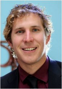
2016 New Investigator: Mark John Hackett

 Nominee:
Nominated By:
Supporting Comments:
What made you choose a career in bioanalysis?
My decision to pursue a career in bioanalysis was influenced by two factors. Firstly, I hold a strong personal interest in the life sciences, specifically neuroscience. Secondly, chemistry was the subject I enjoyed most at school, which led to the decision to study chemistry at University. Although I enjoyed all aspects of training as a chemist, spectroscopic characterization of compounds synthesized in organic and inorganic labs was what I enjoyed most. Thus, the opportunity to undertake research using spectroscopic techniques to quantify chemical markers in biological samples, particularly brain tissue, held strong appeal, and led to my current career path.
Describe the main highlights of your bioanalytical research, and its importance to the bioanalytical community.
During my postgraduate research I became increasingly aware that a niche remained unfilled in the field of analytical neuroscience. Specifically, many important biochemical parameters that can be studied via bulk ex vivo analysis cannot be imaged in situ within tissue sections. Consequently, substantial gaps in knowledge exist in regards to the exact biochemical and physiological pathways that occur during many brain diseases or disorders, hindering therapy development. For example, bulk levels of ions (Cl–, K+, Ca2+), transition metals (Fe, Cu, Zn), small mobile molecules (lactate and taurine), and macromolecular oxidation products (i.e., aggregated proteins, thiol/disulfide ratio) can be detected via bulk chemical assays, but are notoriously difficult to directly image in situ at the cellular level. The main highlight of my research has been to help develop, optimize and validate the use of direct spectroscopic imaging techniques, specifically those that use synchrotron radiation, to fill this niche. The techniques include Fourier transform infrared spectroscopy (FTIR), X-ray absorption spectroscopy (XAS), and X-ray fluorescence microscopy (XFM). A major contribution I made to this field was the optimization and validation of appropriate sample preparation techniques that best preserve the in vivo chemistry of brain tissue, to minimize confounding redistribution or contamination artifacts.
What is the impact of your work beyond your home laboratory?
My PhD research into the optimization and validation of sample preparation protocols for spectroscopic analysis of brain tissue is currently used as sample preparation guidelines by two international synchrotron facilities, the Australian Synchrotron and the Canadian Light Source. The manuscript was published in the Analyst in 2011, and currently has >50 citations.
During my post-doctoral fellowship at the University of Saskatchewan I helped develop and implement single-beam wide-field FTIR imaging at the Canadian Light Source, the first time a method for rapid sub-cellular synchrotron-FTIR imaging has been available in Canada. In addition to my own research, the method was adopted by the Canadian Research Chair for hemorrhagic stroke and the Saskatchewan Research Chairs for Stroke and Multiple Sclerosis. The technique is currently being used by multiple investigators studying various neurological diseases, as well as expanded applications to other biological samples (i.e., cartilage tissue, and plant tissue).
Describe the most difficult challenge you have encountered in the laboratory and how you overcame it.
The greatest challenge in my research has been the development and optimization of sample preparation protocols to allow multiple spectroscopic measurements in combination with routine histology and microscopy to be performed on adjacent tissue sections of the same sample. I overcame this obstacle through several detailed and methodical studies on the specific biochemical parameters that are detected by each spectroscopic method, the limits of detection for each method and the magnitude of biochemical changes that are expected to occur during disease, and the effect of different sample preparation methods on the chemical species detected by each spectroscopic method. Critically, these studies were validated with traditional bulk chemical assays.
This approach enabled my research to quantify the extent to which re-distribution, leaching, contamination and artificial tissue oxidation occur during various sample preparation protocols, and how they can be minimized via rapid cryo-preservation of brain tissue.
The results of this study were in contrast to previous views in the research field, and I believe the acceptance and adoption of the sample preparation protocols I developed could only be achieved through effective communication and dissemination of my research findings, supported by thorough and detailed analytical research methodology and experimental design.
Describe your role in bioanalytical communities/groups.
I strongly believe that the field of bioanalysis can continue to make major contributions to the health research community, which will translate to improved health for society and reduce the economic and emotional burden associated with poor health. For this reason I am actively engaged with the bioanalytical community. I previously served as an academic advisor for the mid infra-red beamline at the Canadian Light Source, where I helped disseminate new imaging advances at the beamline to the health research community; and through engagement with the health research community I helped drive developments at the beamline that would be of benefit to the health research community. I am currently serving on the program advisory committee for the X-ray fluorescence microscopy beamline at the Australian Synchrotron, with a similar role to that described for the Canadian Light Source. In 2012 I was the lead organizer of a PhD and post-graduate one day conference as part of the Canadian Institute of Health Research – Training in Health Research Using Synchrotron Techniques (CIHR-THRUST) program. I am currently a peer reviewer for analytical and interdisciplinary journals: Analyst, Journal of Analytical Atomic Spectrometry, Scientific Reports,Vibrational Spectroscopy.
Please list up to five of your publications in the field of bioanalysis:
- Hackett MJ, Smith SE, Caine S, Nichol H, George GN, Pickering IJ, Paterson PG. Novel bio-spectroscopic imaging reveals altered protein homeostasis and protein aggregation prior to CA1 pyramidal neuron death induced by global brain ischemia in the rat. Free Radicals in Biology and Medicine. 89, 806–818 (2015).
- Hackett MJ, McQuillan JA, El-Assaad F, Aitken JA, Levina A, Cohen D., Siegele R, Carter EA, Grau GE, Hunt NH, Lay PA. Spectroscopic Studies of Molecular and Elemental Alterations in Murine Brain Tissue Induced by Formalin Fixation. Analyst. 136, 2941–2952 (2011).
- Hackett MJ, Aitken JB, El-Assaad F, McQuillan JA, Carter EA, Ball HJ, Tobin MJ, Paterson D, de Jonge MD, Siegele R, Cohen DD, Vogt S, Grau GE, Hunt NH, Lay PA. Mechanisms of murine cerebral malaria: Multimodal imaging of altered cerebral metabolism and protein oxidation at hemorrhage sites. Science Advances doi:10.1126/sciadv.1500911 (2015).
- Hackett MJ, Smith SE, Paterson PG, Nichol H, Pickering IJ, George GN. X-ray Absorption Spectroscopy at the Sulfur K-Edge: A New Tool to Investigate the Biochemical Mechanisms of Neurodegeneration. ACS Chemical Neuroscience. 3, 178–188 (2012).
- Hackett MJ, DeSouze M, Caine S, Nichol H, Paterson PG, Colbourne F. A new method to image heme-Fe, total Fe and aggregated protein levels after intra-cerebral haemorrhage. ACS Chemical Neuroscience. 6, 761–770. (2015)
Please select one publication from above that best highlights your career to date in the field of bioanalysis and provide an explanation for your choice (100 word limit)
Publication 1 best highlights my career achievements to date, as it incorporates single-beam wide-field FTIR imaging that I helped develop at the Canadian Light Source, and the results could not have been obtained without the use of cryo-preserved brain tissue. The results of the study revealed increased protein aggregation within a distinct population of hippocampal neurons after brain ischemia (i.e., stroke), prior to altered lipid homeostasis and cell degeneration. Due to the large memory deficits associated with stroke and many neurodegenerative diseases, this study suggests further research into protein aggregation at early disease time points may identify novel therapeutic strategies.
Find out more about this year’s New Investigator Award, the prize, the judging panel and the rest of our nominees.


